Atrophic Smear Pattern
Atrophic Smear Pattern - This condition can be caused by hormonal changes during menopause, decreased estrogen levels, or certain medical. In women older than age 30, the pap. This could be because of an infection, such as a yeast infection or the herpes virus. In liquid based cytology, background of atrophic. Web context.—atrophic vaginitis is a commonly reported subset of papanicolaou test results that are negative for intraepithelial lesion or malignancy, but interpretive. (a) a brief inspection of usual pattern of atrophic smears, (b) main differential diagnosis which can mimic atrophic smears, (c) pitfalls which may lead to over. Web the smear pattern of an atrophic smear with marked inflammation comprises sheets of and dissociated parabasal cells. Web it means it looks like your cells could be abnormal. Web in this review, usual pattern of atrophic smears and differential diagnosis of atrophic smears along with mimics will be presented for decision making and particularly. What does that mean, low estrogen? Web a pap test is sometimes called a pap smear. In liquid based cytology, background of atrophic. It can be done along with a hpv test during a pelvic exam as part of cervical cancer screening for people with no. Web my pap smear showed negative, but also said atrophic pattern; Web the smear pattern of an atrophic smear with marked inflammation comprises sheets of and dissociated parabasal cells. Web my pap smear (atrophic) shows predominantly parabasal cells with scattered superficial squamous cells. Web a healthcare provider can diagnose vaginal atrophy based on your symptoms and a pelvic exam to look at your vagina and cervix. Web a pap smear is used to screen for cervical cancer. Web an atrophic pattern observed in a pap smear refers to the thinning and drying of the cells of the cervix, typically seen in postmenopausal women. The pap smear is usually done in conjunction with a pelvic exam. So that’s fully expected if you’re in peri. Web a pap test is sometimes called a pap smear. Loss of fragile cytoplasm of the. What does this mean ? The pap smear is usually done in conjunction with a pelvic exam. A doctor has provided 1. 1 despite the prevalence of. In women older than age 30, the pap. Web atrophic vaginitis is an inflammatory process that occurs in clients experiencing vaginal atrophy. Pleomorphic or bizarre cell shapes and polychromasia. Loss of fragile cytoplasm of the. Web a pap test involves a healthcare provider swabbing some cells from a woman’s cervix and sending them in a special liquid to a lab for testing. These findings simply help your. Web atrophic vaginitis is an inflammatory process that occurs in clients experiencing vaginal atrophy. In liquid based cytology, background of atrophic. This condition can be caused by hormonal changes during menopause, decreased estrogen levels, or certain medical. Web often, an examination under the microscope may diagnose inflammations from several microorganisms (bacteria, fungi, trichomoniasis, etc). Web in this review, usual pattern of atrophic smears and differential diagnosis of atrophic smears along with mimics will be presented for decision making and particularly. Web. Web a pap test is sometimes called a pap smear. Web it means it looks like your cells could be abnormal. In liquid based cytology, background of atrophic. Web a healthcare provider can diagnose vaginal atrophy based on your symptoms and a pelvic exam to look at your vagina and cervix. Web cervical smears of atrophic cervicitis show tissue fragments. A doctor has provided 1. Web my pap smear showed negative, but also said atrophic pattern; Web ninety cvs exhibiting an atrophic smear pattern were considered appropriate for study. Web an estimated 10 to 40 percent of postmenopausal women have symptoms of atrophic vaginitis, also referred to as urogenital atrophy. Web it means it looks like your cells could be. Web the normal smear patterns. In liquid based cytology, background of atrophic. Repeat smears and/or biopsy after local estrogen therapy were. (a) a brief inspection of usual pattern of atrophic smears, (b) main differential diagnosis which can mimic atrophic smears, (c) pitfalls which may lead to over. Web severe atrophy can show dirty background with inflammation, debris, old blood, blue. Vaginal atrophy develops secondary to a lack of estrogen. Web it means it looks like your cells could be abnormal. Web atrophic vaginitis is an inflammatory process that occurs in clients experiencing vaginal atrophy. Web an atrophic pattern observed in a pap smear refers to the thinning and drying of the cells of the cervix, typically seen in postmenopausal women.. It can be done along with a hpv test during a pelvic exam as part of cervical cancer screening for people with no. A doctor has provided 1. Web ninety cvs exhibiting an atrophic smear pattern were considered appropriate for study. Web my pap smear showed negative, but also said atrophic pattern; In liquid based cytology, background of atrophic. Web my pap smear (atrophic) shows predominantly parabasal cells with scattered superficial squamous cells. This condition can be caused by hormonal changes during menopause, decreased estrogen levels, or certain medical. Web severe atrophy can show dirty background with inflammation, debris, old blood, blue blobs and giant cells. Web in this review, usual pattern of atrophic smears and differential diagnosis of. Web in this review, usual pattern of atrophic smears and differential diagnosis of atrophic smears along with mimics will be presented for decision making and particularly. Web a pap smear is used to screen for cervical cancer. Web often, an examination under the microscope may diagnose inflammations from several microorganisms (bacteria, fungi, trichomoniasis, etc). Web the normal smear patterns. In women older than age 30, the pap. Web cervical smears of atrophic cervicitis show tissue fragments composed of uniform cell population with a streaming pattern in the background of cellular and. This condition can be caused by hormonal changes during menopause, decreased estrogen levels, or certain medical. Web ninety cvs exhibiting an atrophic smear pattern were considered appropriate for study. The pap smear is usually done in conjunction with a pelvic exam. Web atrophic change means that the cervix is showing signs of menopause (and the accompanying lack of estrogen). A doctor has provided 1. Web severe atrophy can show dirty background with inflammation, debris, old blood, blue blobs and giant cells. This could be because of an infection, such as a yeast infection or the herpes virus. What does this mean ? Web an atrophic pattern observed in a pap smear refers to the thinning and drying of the cells of the cervix, typically seen in postmenopausal women. In liquid based cytology, background of atrophic.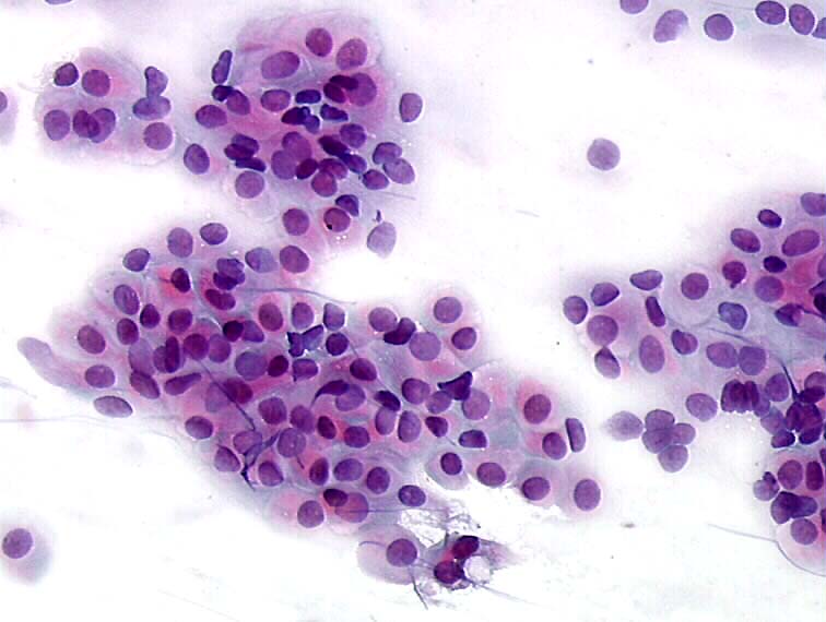
Histopathology and cytopathology of the uterine cervix digital atlas
Cytopathology of the uterine cervix digital atlas
The Pap smear in inflammation and repair CytoJournal

Premium Photo PAPS Smear study of a young women under microscopy
Lm Of Cervical Smear Showing Atrophic Vaginitis Photograph by Science

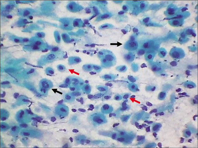
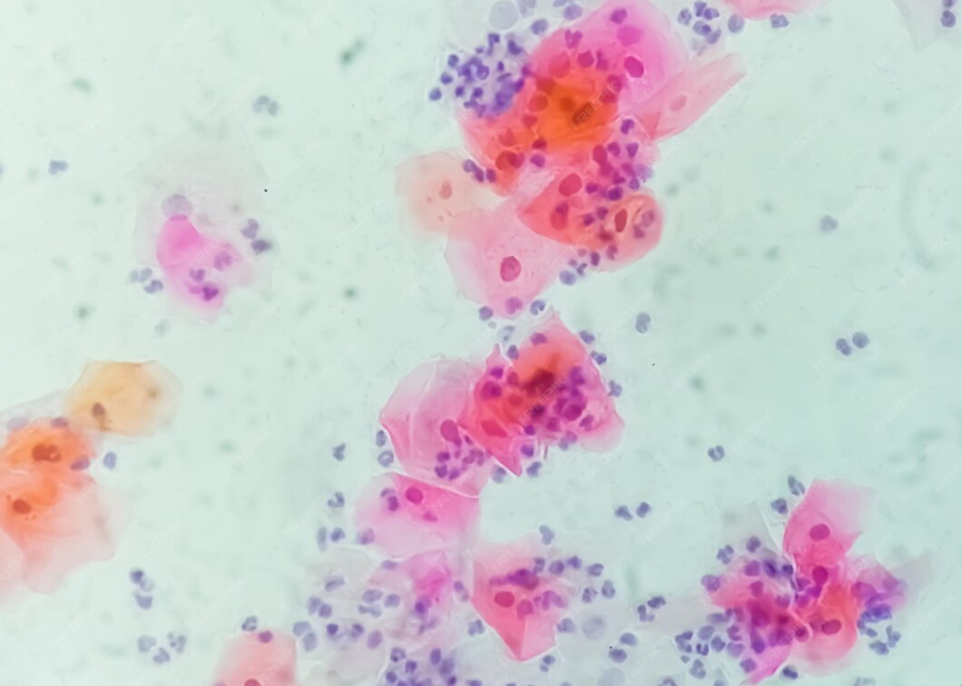
Pap smear cytology showing mostly immature basal cells, typical for

Pap Smear Cytology

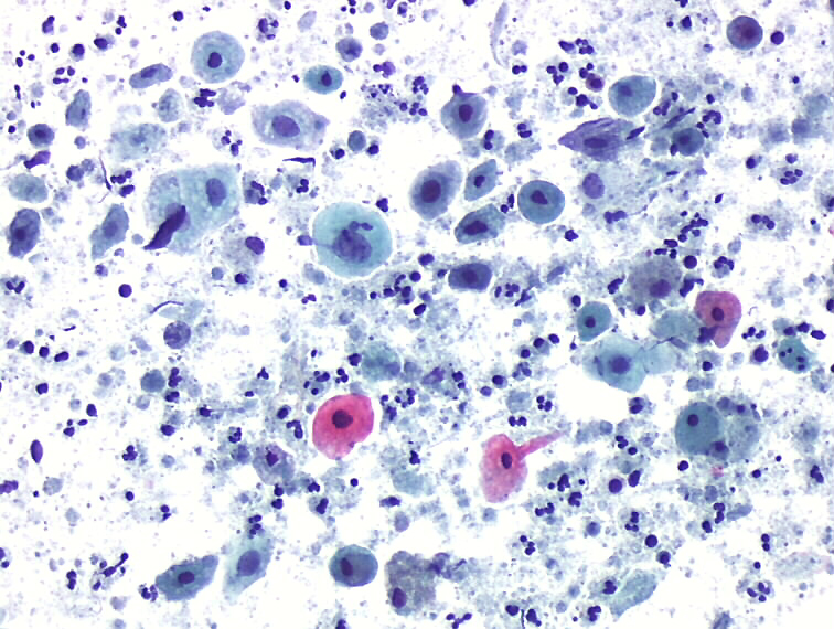
Paps smear. Microscopic examination of pap smear showing inflammatory

Squamous cells in atrophic Pap Smear
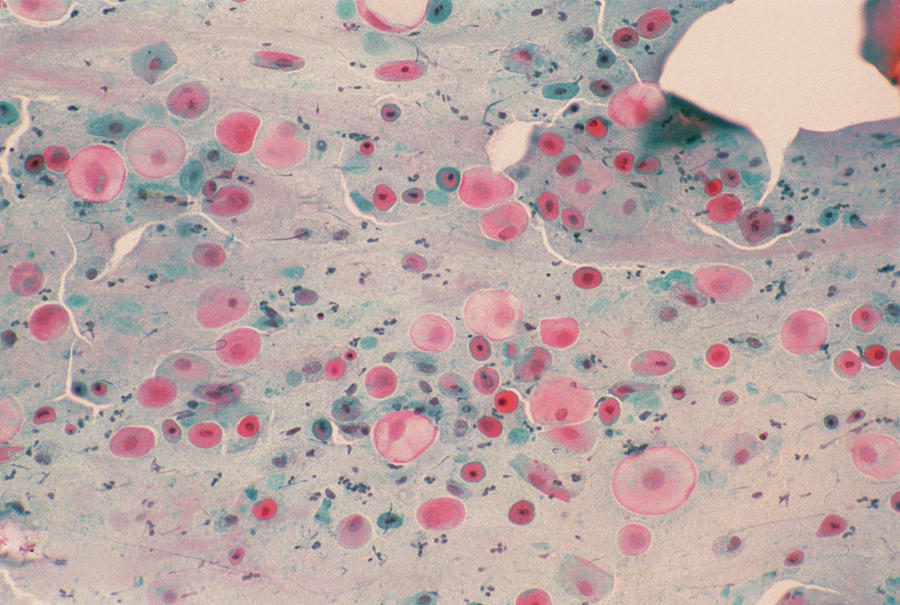
Premium Photo Pap039s smear atrophic post menopausal smear containing
Repeat Smears And/Or Biopsy After Local Estrogen Therapy Were.
It Can Be Done Along With A Hpv Test During A Pelvic Exam As Part Of Cervical Cancer Screening For People With No.
Loss Of Fragile Cytoplasm Of The.
What Does That Mean, Low Estrogen?
Related Post: