Flow Volume Loop Patterns
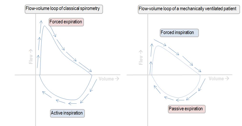
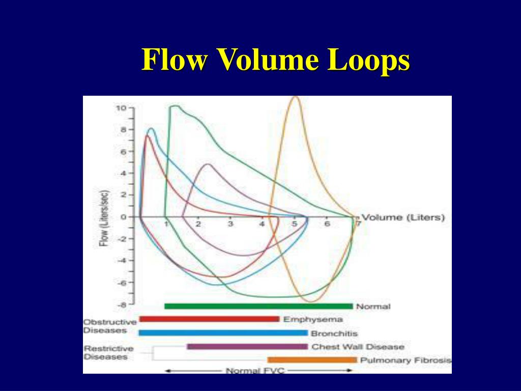
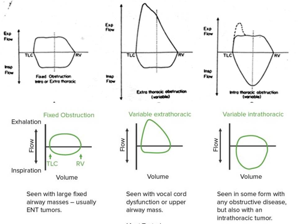
Flow Volume Loop Patterns - Web the flow volume loop suggested that the nonobstructed side of the lung empties rapidly, and the obstructed side empties slowly throughout the forced vital capacity maneuver. The airways are divided into intrathoracic and extrathoracic components by the thoracic inlet. Web flow volume loops. Consequently, the expiratory portion of the flow volume loop demonstrates both a restrictive pattern and a severe obstructive pattern in series and is unlike any of the. It demonstrates the obstructive, restrictive and mixed pattern lung pathology. Web a rounded peak flow can be an indication of an inadequate subject effort. An abrupt drop in expiratory flow usually indicates an early termination of exhalation. Web airflow and lung volume measurements can be used to differentiate obstructive from restrictive pulmonary disorders, to characterize severity, and to measure responses to therapy. Low maximum forced expiratory flow, biphasic expiratory curve, flow oscillations, and notching. Changes in the contour of the loop can aid in the diagnosis and localization of airway obstruction [ 1 ]. Changes in the contour of the loop can aid in the diagnosis and localization of airway obstruction [ 1 ]. An abrupt drop in expiratory flow usually indicates an early termination of exhalation. Web ventilator waveforms are graphical descriptions of how a breath is delivered to a patient. Web a rounded peak flow can be an indication of an inadequate subject effort. This finding has also been described as “two compartments.” a ct chest was obtained which showed an obstructing right mainstem bronchus lesion. Web the flow volume loop suggested that the nonobstructed side of the lung empties rapidly, and the obstructed side empties slowly throughout the forced vital capacity maneuver. Failure of the expiratory flow curve to reach zero (an open loop) a scooped out expiratory flow pattern. Obstruction of the large airways. Changes in the contour of the loop can aid in the diagnosis and localization of airway obstruction [ 1 ]. Web a restrictive pattern is indicated by an fvc below the fifth percentile based on nhanes iii data in adults, or less than 80% in patients five to 18 years of age. Web airflow and lung volume measurements can be used to differentiate obstructive from restrictive pulmonary disorders, to characterize severity, and to measure responses to therapy. It demonstrates the obstructive, restrictive and mixed pattern lung pathology. Provide a graphical analysis of inspiratory and expiratory flow from various inspired lung volumes. Changes in the contour of the loop can aid in the. Web ventilator waveforms are graphical descriptions of how a breath is delivered to a patient. Low maximum forced expiratory flow, biphasic expiratory curve, flow oscillations, and notching. Consequently, the expiratory portion of the flow volume loop demonstrates both a restrictive pattern and a severe obstructive pattern in series and is unlike any of the. An abrupt drop in expiratory flow. Changes in the contour of the loop can aid in the diagnosis and localization of airway obstruction [ 1 ]. Web flow volume loops. The airways are divided into intrathoracic and extrathoracic components by the thoracic inlet. Provide a graphical analysis of inspiratory and expiratory flow from various inspired lung volumes. This finding has also been described as “two compartments.”. Low maximum forced expiratory flow, biphasic expiratory curve, flow oscillations, and notching. Failure of the expiratory flow curve to reach zero (an open loop) a scooped out expiratory flow pattern. Web airflow and lung volume measurements can be used to differentiate obstructive from restrictive pulmonary disorders, to characterize severity, and to measure responses to therapy. Changes in the contour of. Changes in the contour of the loop can aid in the diagnosis and localization of airway obstruction [ 1 ]. An abrupt drop in expiratory flow usually indicates an early termination of exhalation. Obstruction of the large airways. It can also indicate unusual abnormalities, such as obstructive lesions of the central airways. Consequently, the expiratory portion of the flow volume. Changes in the contour of the loop can aid in the diagnosis and localization of airway obstruction [ 1 ]. This finding has also been described as “two compartments.” a ct chest was obtained which showed an obstructing right mainstem bronchus lesion. It can also indicate unusual abnormalities, such as obstructive lesions of the central airways. Web a restrictive pattern. Failure of the expiratory flow curve to reach zero (an open loop) a scooped out expiratory flow pattern. Web ventilator waveforms are graphical descriptions of how a breath is delivered to a patient. Web a restrictive pattern is indicated by an fvc below the fifth percentile based on nhanes iii data in adults, or less than 80% in patients five. Changes in the contour of the loop can aid in the diagnosis and localization of airway obstruction [ 1 ]. It can also indicate unusual abnormalities, such as obstructive lesions of the central airways. Provide a graphical analysis of inspiratory and expiratory flow from various inspired lung volumes. Failure of the expiratory flow curve to reach zero (an open loop). This finding has also been described as “two compartments.” a ct chest was obtained which showed an obstructing right mainstem bronchus lesion. Web airflow and lung volume measurements can be used to differentiate obstructive from restrictive pulmonary disorders, to characterize severity, and to measure responses to therapy. Web ventilator waveforms are graphical descriptions of how a breath is delivered to. Low maximum forced expiratory flow, biphasic expiratory curve, flow oscillations, and notching. Obstruction of the large airways. It demonstrates the obstructive, restrictive and mixed pattern lung pathology. Consequently, the expiratory portion of the flow volume loop demonstrates both a restrictive pattern and a severe obstructive pattern in series and is unlike any of the. The airways are divided into intrathoracic. Obstruction of the large airways. This finding has also been described as “two compartments.” a ct chest was obtained which showed an obstructing right mainstem bronchus lesion. Consequently, the expiratory portion of the flow volume loop demonstrates both a restrictive pattern and a severe obstructive pattern in series and is unlike any of the. Web flow volume loops. Changes in the contour of the loop can aid in the diagnosis and localization of airway obstruction [ 1 ]. Low maximum forced expiratory flow, biphasic expiratory curve, flow oscillations, and notching. Changes in the contour of the loop can aid in the diagnosis and localization of airway obstruction [ 1 ]. Web ventilator waveforms are graphical descriptions of how a breath is delivered to a patient. Web a restrictive pattern is indicated by an fvc below the fifth percentile based on nhanes iii data in adults, or less than 80% in patients five to 18 years of age. Web airflow and lung volume measurements can be used to differentiate obstructive from restrictive pulmonary disorders, to characterize severity, and to measure responses to therapy. Web the flow volume loop suggested that the nonobstructed side of the lung empties rapidly, and the obstructed side empties slowly throughout the forced vital capacity maneuver. Failure of the expiratory flow curve to reach zero (an open loop) a scooped out expiratory flow pattern. Web a rounded peak flow can be an indication of an inadequate subject effort. It demonstrates the obstructive, restrictive and mixed pattern lung pathology.
Pulmonary FlowVolume Loops & Disease Medical science, Pathology

Pin on Pulmonary

Flow Volume Loops Common Patterns A. Normal B. GrepMed
Interpreting the shape of the flowvolume loop Deranged Physiology
Interpreting the shape of the flowvolume loop Deranged Physiology
PPT Pulmonary Function Tests Jonathan Kass PowerPoint Presentation
Flow Volume Loops Explained

Flow Volume Loops Respiratory Physiology Pulmonary Medicine

Kyphoscoliosis Flow Volume Loop

Pulmonary Function Tests Pulmonary Medbullets Step 2/3
The Airways Are Divided Into Intrathoracic And Extrathoracic Components By The Thoracic Inlet.
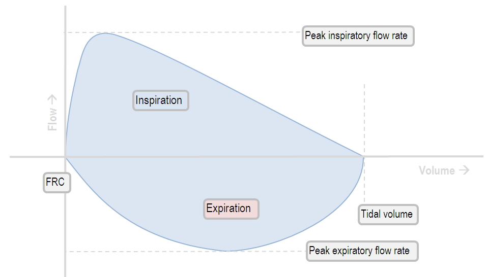
Provide A Graphical Analysis Of Inspiratory And Expiratory Flow From Various Inspired Lung Volumes.
An Abrupt Drop In Expiratory Flow Usually Indicates An Early Termination Of Exhalation.
It Can Also Indicate Unusual Abnormalities, Such As Obstructive Lesions Of The Central Airways.
Related Post: